

Researchers at the University of Regensburg and doctors at the university hospital are developing a novel screening tool to improve tumor treatments
20.03.2023 15:51
According to the Robert Koch Institute, around 19,000 people develop pancreatic carcinoma every year. The five-year survival rate is only 8-10%, making pancreatic carcinoma the fourth most common cause of death among all types of cancer. For this reason, it is important to obtain new insights through research in order to be able to develop new therapy methods.
Metastatic pancreatic carcinoma in particular is considered incurable. In this case, the tumor has already released cells into the blood that can form metastases elsewhere in the body. A quick diagnosis and a quick start to therapy are extremely important here. After the tumor is surgically removed, patients receive adjuvant chemotherapy to destroy tumor cells that are still in the body. Nevertheless, pancreatic carcinoma often develops resistance to treatment. There are also targeted, personalized therapy procedures that influence important tumor signaling pathways. Despite the different treatment methods, there has been no significant breakthrough in the treatment of pancreatic carcinoma in the last 30 years.
The working groups of Prof. Dr. Silke Härteis (Chair of Molecular and Cellular Anatomy, Univ. Regensburg), Prof. Dr. Thiha Aung (Chair of Molecular and Cellular Anatomy, Univ. Regensburg; Deggendorf University of Technology), Prof. Dr. Christoph Brochhausen-Delius (formerly the Institute of Pathology at the UR, now Director of the Pathological Institute at the University Hospital Mannheim) and Prof. Dr. Christina Hackl (Clinic and Polyclinic of Surgery, Regensburg University Hospital) are dedicating themselves to this important topic in a research project that is part of the “Central Innovation Program for SMEs” funding program of the Federal Ministry for Economic Affairs and Climate Protection (BMWK; project form R&D cooperation projects; AiF Projekt GmbH). ¾ million euros will be funded.
Development of a drug screening platform
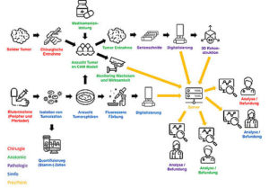
The project is developing a novel screening tool: a drug screening platform. This will make it possible to determine the effect of the therapeutic agents on the tumors at every treatment period in order to then be able to carry out the best possible therapy. For this purpose, the scientists from the university and the clinic have joined forces with other cooperation partners from the clinic and industry to combine various innovative methods.
In order to obtain samples for the project, patients at the University Hospital Regensburg are informed about the planned treatment and the possibility of taking part in the study. Ms. Prof. Dr. Christina Hackl (Clinic and Polyclinic of Surgery, Regensburg University Hospital) coordinates the study-related process within the surgical department. If a patient agrees to participate, blood and tumor samples that are not needed for diagnostic purposes can be brought to the project partner immediately following preoperative or surgical collection.
The project partner simfo GmbH from Bayreuth isolates the subpopulation of tumor cells from the blood of patients in order to grow so-called tumor spheres (spherical cell colonies) from the cells. The company simfo GmbH already has many years of experience in the morphological, molecular biological and functional characterization of cells from solid epithelial tumors released into the blood.
In the working group of Prof. Dr. Härteis and Prof. Dr. The resulting tumor spheres and the primary material of the pancreatic tumor are then cultured using the chorionic allantoic membrane model (CAM model). The tumor spheres and the tumor tissue are grown on the CAM membrane as so-called “patient-derived xenografts” (PDX), which nourish the tumor cells and the tumor tissue like a placenta.
Evaluate and characterize tumor growth at any time
Thanks to the experimental setup, tumor growth can be evaluated at any time. In this way, the effectiveness and selectivity of established and new therapeutic agents on the growth of tumor cells as well as on angiogenesis processes (formation of new blood vessels) and the ingrowth of the tumor into the surrounding tissue can be analyzed directly. Resistance to therapeutic agents can also be detected in this way.
The exact characterization of the tumor tissue and the tumor cells takes place in part in the Institute of Pathology in the working group of Prof. Dr. Brochhausen-Delius through structural and ultrastructural analyses. There, histological, immunohistological and electron microscopic examinations are carried out on the tumor spheres and tumor tissue. For this purpose, sectional preparations are created using the different samples from the CAM model. The samples are scanned with the M8 microscope (PreciPoint GmbH) in order to produce overview images of the sections, so-called “Whole Slide Images” (WSI), for the analyses. With the help of serial sections, a 3D reconstruction of the immunohistologically stained blood vessels can also be carried out in order to show the vascular system and its branches more precisely. The analyzes can ultimately be used to make statements about functional molecules, growth of the tumor and blood vessels in order to gain a deeper understanding of the blood supply to the tumor and its structure, also in comparison to metastatic tumors.
Another part of the histological and molecular biological analyzes takes place in the laboratory of Prof. Dr. Hackl, who also carries out a quantitative evaluation of proliferation ability, angioinvasion, tissue invasion and response to chemotherapy. At the same time, Prof. Hackl provides continuous follow-up care for patients during surgical oncology consultations and correlates the clinical course with the tumor characteristics collected by the project.
The industrial partner PreciPoint GmbH from Freising will create an online platform, thereby enabling the virtual networking of surgery, laboratory medicine, anatomy, pathology and basic research. With the help of the platform, metadata, diagnostic data and image data can be shared directly, allowing different departments to work together quickly and effectively. The development of new algorithms enables structural analyzes of the data and active ingredient screening of samples with certain characteristics. Another innovation will be a combination of brightfield and fluorescence-based microscopy to make it easier to display the images and view them between different acquisition modes.
The ultimate goal of the drug screening platform is to determine the best treatment option with the fewest side effects within a very short period of time for the affected patient in order to improve the treatment of pancreatic carcinoma and ultimately other tumors in the future.